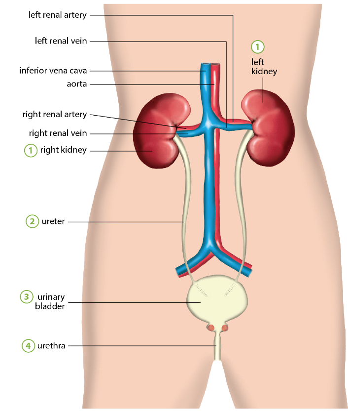
 Consists of a pair of kidneys, a pair of ureters, urinary bladder and urethra
| Part | Elaboration |
|---|---|
| Kidneys Structure of the Kidney | ⇒ beaned shaped organs ⇒ embedded in a mass of fat in the abdominal cavity Average size ⇒ of the human kidney is 10-13cm long and about 5-7cm wide - attached to the dorsal body wall with one on each side of the vertebral column - concave surface faces the vertebral column. - depression at the centre of the concave surface (hilus) - Renal artery, renal vein, and nerves are connected to the kidneys through the hilus - made up of nephrons ⇒ lie just above the waistline left kidney is slightly higher than the right kidney main function of Kidneys: Excrete urea and excess salt and water as urine |
| Ureter | Narrow tube ⇒ emerges from hilus ⇒ travels downwards to join the dorsal surface of the urinary bladder (connects kidney to urinary bladder) urine from each kidney > passes through the ureter to the urinary bladder |
| Urinary Bladder | → elastic muscular bag located in front of the rectum → stores urine - ventral to rectum - bottom of urinary bladder is the sphincter muscle - sphincter muscle relaxes when bladder is full → allowing urine to flow into urethra |
| Urethra | → duct through which urine passes from the bladder to outside of the body females (4cm) males (about 20cm) |