| Implantation |
|---|
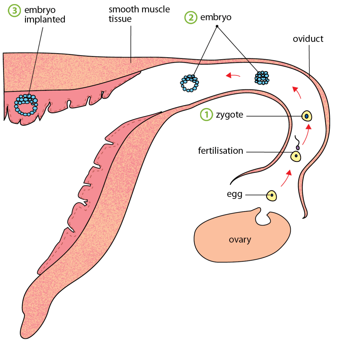
| 1. Cilia lining the inner surface of oviduct → sweep fertilised egg/zygote → along oviduct to the uterus (at the same time mitosis occurs, refer to step 2.) 2. zygote → divides by mitosis → to form hollow ball of cells → embryo. Energy needed for this early stage of cell division is provided by the nutrients stored in the egg. Takes about 5 days for embryo to reach uterus. Embryo may float freely in the uterus for about 2 days. 3. Eventually → embryo sinks into / becomes embedded in the uterine lining. Called implantation. Implantation usually occurs abt 7 days after fertilisation. |
 |
| Development of Placenta | Development of Amniotic Sac |
|---|---|
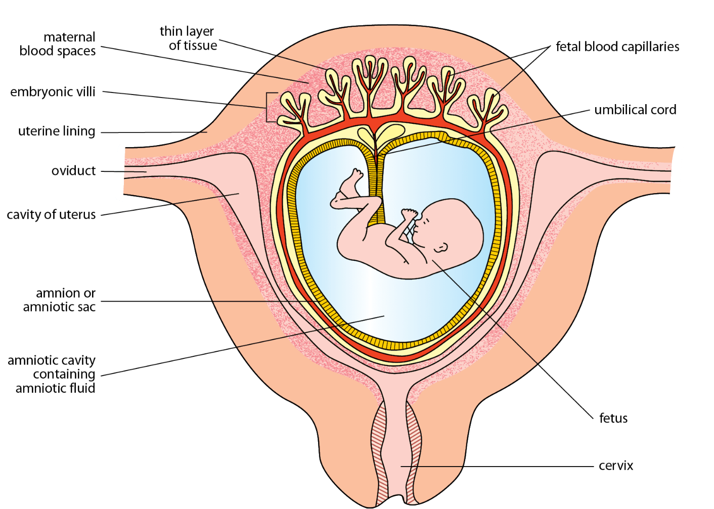
| 4. soon after implantation → finger-like projections → villi → begin to grow from embryo → into uterine lining. They contain blood capillaries of embryo. 5. villi + uterine lining → in which villi are embedded → make up the placenta 6. tube → known as umbilical cord → attaches the embryo to the placenta | 7. membrane called amniotic sac or amnion → begins to develop about same time as placenta 8. amniotic sac → encloses embryo in fluid filled space → amniotic cavity. Fluid in amniotic cavity is called amniotic fluid 9. embryo continues to develop. in humans, abt 10-12 weeks after fertilisation → all major organs are formed. from this stage onwards, the embryo is known as fetus |
| ![[Pasted image 20250410100625.png | 500]] |
Functions of Placenta
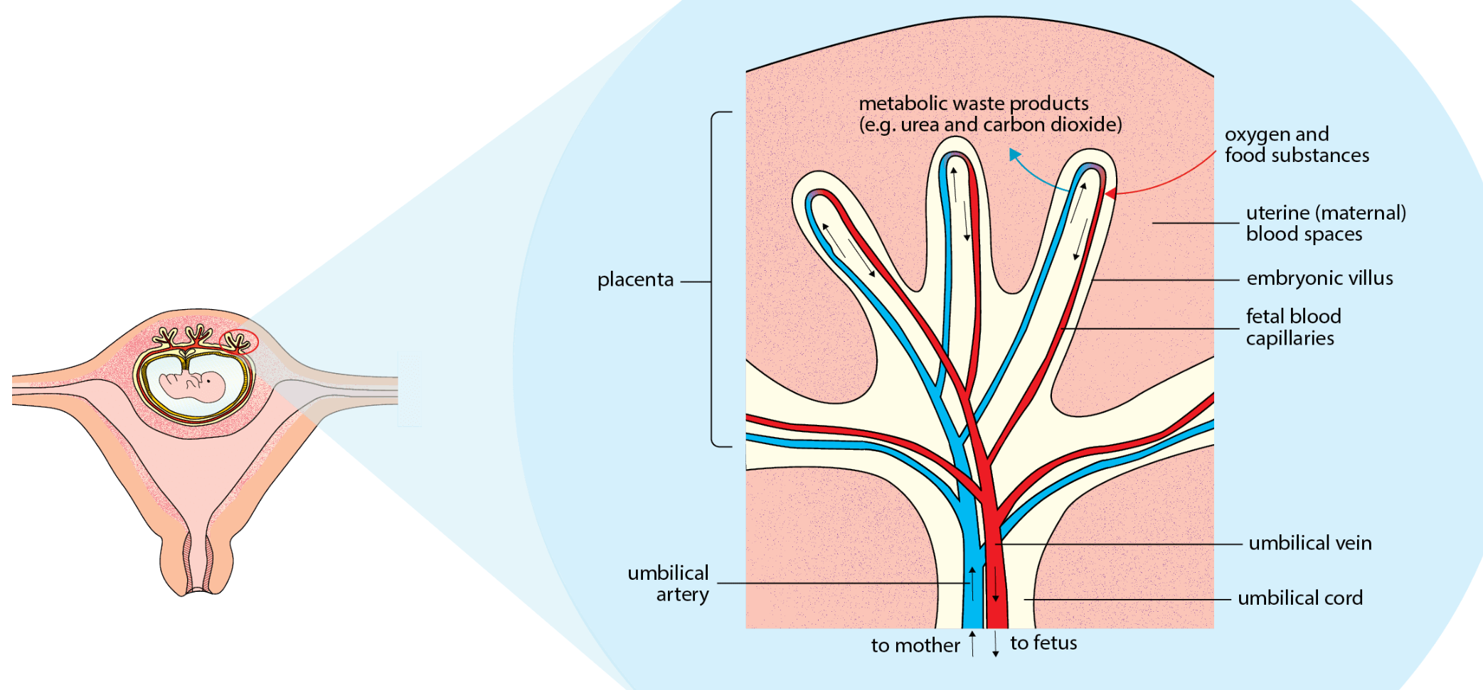
- allows oxygen + dissolved food substances → such as glucose, amino acids, mineral salt → to diffuse from maternal blood into fetal blood.
- allows metabolic waste / excretory products → such as urea and carbon dioxide → to diffuse from fetal blood → into maternal blood.
- allows protective antibodies → to diffuse from maternal blood → into fetal blood. antibodies protects fetus → against certain diseases.
- produces oestrogen and progesterone which maintain uterine lining in a healthy state during pregnancy.
Functions of Umbilical Cord
attaches fetus to placenta. umbilical cord → contains blood vessels of the fetus.
Contains:
- 2 umbilical arteries → transport deoxygenated blood and metabolic waste products → from fetus to placenta
- babies with 1 artery are usually smaller.
- one umbilical vein → that transported oxygenated blood and food substances from placenta to fetus
Functions of Amniotic Fluid
amniotic sac → encloses fetus → in amniotic cavity → which contains amniotic fluid.
functions:
- supports and cushions fetus → before birth. it is a shock absorber. as it cannot be compressed → it protects fetus against physical/mechanical injury.
- buoys up fetus → allow\ fetus → a certain degree of movement → which promotes muscular development.
- during birth → it lubricates + reduces fiction → in vagina / birth canal
Extra: Amniotic fluid keeps fetus at correct temperature. Not lower than mothers temperature*
Fetal blood system is Separated from Maternal Blood System
- fetal blood capillaries → surrounded by maternal blood spaces.
- in placenta → fetal blood capillaries are separated from maternal blood system → only by thin layer of tissue
- diffusion of dissolved substances → can occur across this layer of tissue
however → pathogens and toxins → that may be present in the maternal bloodstream → can pass across placenta and affect the fetus.
Why is Fetal Blood System Not Continuous with Maternal Blood System
feternal blood system is not continuous with the maternal blood system
2 blood systems cannot be continuous because:
- blood pressure of mother → would kill fetus → as it is much higher than that of fetus
- blood group of fetus → may not be same as mothers. if fetus and mother have diff blood groups and 2 systems allowed to mix → antibodies in mothers group may cause fetal blood cells to agglutinate. dangerous to both mother and baby
- prevents potential harmful substances or pathogens from directly entering fetal bloodstream.