Simple unicellular organisms → No part of the cell is far from external environment.
- O2 and nutrients → can diffuse through cell membrane and easily reach all parts of the cell.
- waste products → can be rapidly removed from cell through diffusion.
complex multicellular organisms → numerous cells are found deep in the body → Far from external environment.
- simple diffusion → cannot bring enough O2 and food materials to these cells in time
- also cannot remove waste products → with sufficient speed. Must be a transport system to carry substances from one part of the body to the other.
circulatory system consists of:
- series of blood vessels that run through the entire body
- blood - which flows through vessels carrying materials through the body
- heart (muscular pump) → ensures fluid keeps flowing through the vessels.
Blood
Blood in this case is an individual topic, so refer to the following page. Blood
How does Blood Clot
Blood exposed to air → will clot or coagulate. clot → seals the wound → preventing excessive loss of blood. Clot → also prevents foreign particles from entering the bloodstream.
People suffering from a disease called Hemophilia → Normal blood clotting mechanism is greatly impaired. slight injuries → if not immediately treated → may cause person to bleed to death → or die of internal bleeding.
Mechanism of Clotting:
- Platelets: and damaged tissues release an enzyme called Thrombokinase
- Thrombokinase catalyses reaction of soluble protein fibrinogen to insoluble fibrin threads.
- Insoluble fibrin threads form a network/mesh which entangles and traps red blood cells.
- the whole mass forms a clot or a scab. the clot seals the wound to prevent entry of micro-organisms and pathogens and bacteria into the bloodstream, as well as prevent excessive loss of blood.
Important
platelets produce an enzyme that catalyses reaction of soluble fibrinogen to insoluble fibrin threads
Sample
When injured → It catalyzes the conversion of prothrombin to thrombin in the presence of calcium ions. thrombin then catalyses conversion of soluble protein fibrinogen to insoluble fibrin threads insoluble fibrin threads then form a network/ mesh which entangles and traps RBC. CONTINUE WITH ABOV EXPLANATIOn
wound incurred Thrombokinase → produced by platelets and wound Blood clots to prevent further loss of blood.
Organ Transplant and Tissue Rejection
Sometimes when persons tissue or organ is damaged or diseased → it can be replaced with a healthy tissue or organ from a donor. → called tissue or organ transplant.
Organ to be transplanted → must not be rejected by recipients immune system. → Recipients White Blood cells→ may respond → by producing antibodies to destroy the transplanted organ.
Similarly foreign tissue → may be recognized by white blood cells. → white blood cells → respond → by producing Antibodies to destroy the foreign tissue. → known as tissue rejection → tissue rejection will not be a problem if tissue to be transplanted comes from the same person
if tissue to transplant comes from a donor → then tissue match s necessary to reduce risk of rejection. → tissues of both donor and recipient must be genetically similar as possible. → another way to reduce risk of tissue rejection is the use of drugs which inhibit the responses of the recipients immune system.
- Lymphocytes(which falls under White Blood cells produce antibodies → to destroy transplanted organs
- Prevention of rejection:
- tissue match (self, family members, close relatives)
- immunosuppressive drugs
- X-ray radiation.
Blood Groups
→ death caused by blood transfusion → due to red blood cells of donor agglutinating in the recipients body. → Such clumps blocked up small blood vessels and obstructed the flow of blood.
4 Different Blood groups
→ surfaces of RBC → contain special proteins → called antigens.
these same antigens → are found on all red blood cells.
Blood plasma → contains antibodies → which are produced by White Blood cells and are always present in blood.
These natural antibodies → will not react with the antigens on your blood cells → but may react with the antigens on RBC of another person.
→ causing clumping of RBC (agglutination)
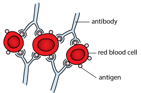 classification of blood groups:
- antigens are represented by capital letters A and B
- Antibodies against antigen A and B → represented by small letters a and b respectively.
*blood group table
| Blood Group | A | B | C | D |
|---|---|---|---|---|
| Red Blood cell | 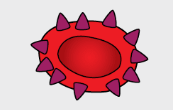 | 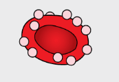 | 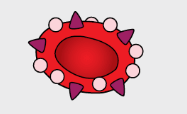 | 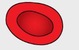 |
| Antigen on Red blood cell | Antigen A | Antigen B | Antigen A and B | No Antigens |
| Antibody in Plasma | Antibody B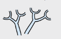 | Antibody A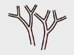 | No Antibodies | Antibodies A and B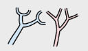 |
| → if you belong to blood group A, your RBC possesses antigen A and plasma contains antibody b. | ||||
| → plasma could not contain antibody a, or you own red blood cells will agglutinate. |
what happens when different blood groups mix?
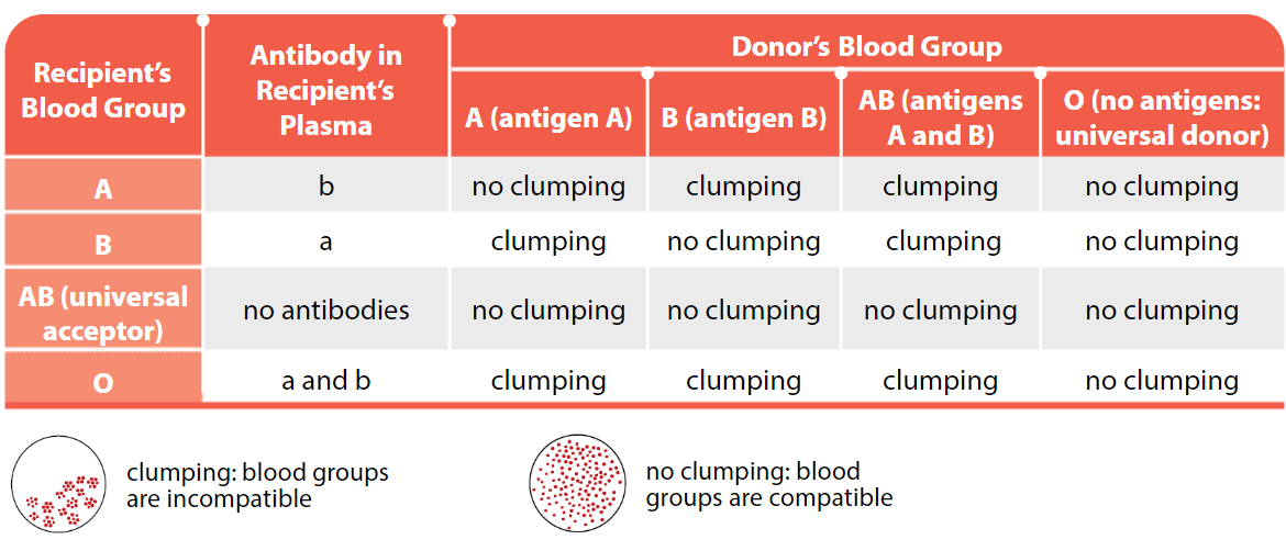 when you donate blood → it is usually separated into its components.
most of the time → only the donors red blood cells are transfused to the recipients.
Red blood cells of group O → do not have any antigens A and B
Hence → when group O red blood cells are transfused into a person of blood group A → recipients antibodies will not agglutinate with donors red blood cells.
→ this is why the person of blood group O → is called universal donor.
when you donate blood → it is usually separated into its components.
most of the time → only the donors red blood cells are transfused to the recipients.
Red blood cells of group O → do not have any antigens A and B
Hence → when group O red blood cells are transfused into a person of blood group A → recipients antibodies will not agglutinate with donors red blood cells.
→ this is why the person of blood group O → is called universal donor.
Functions of Blood;
Functions of Blood (individual topic)
Parts of the Circulatory System
- Blood → used to transport various substances from one part of the body to another by flowing continuously around the body.
- In vertebrates → blood flows through a close system of blood vessels called circulatory system.
- this blood flow → called blood circulation
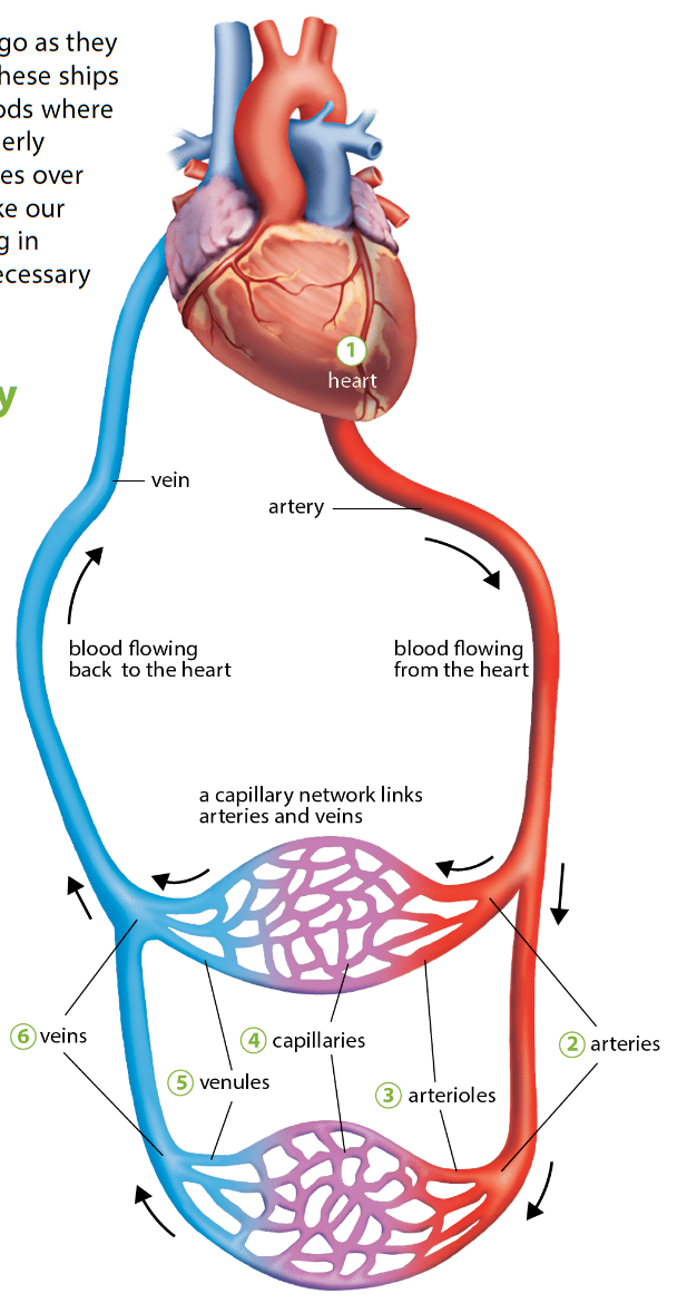
Overview of the parts of the Circulatory system
-
heart Blood → kept circulating throughout the body by means of a muscular pump → heart. When heart relaxes → it fills up with blood. When it contracts → blood is squeezed/forced out with a great force (DO NOT SAY PUMPED) Blood → then circulates through blood vessels → which direct flow of blood throughput the body.
-
Arteries Blood vessels that carry blood away from the heart Large artery that leaves the left side of the heart → Aorta. Branches out to form smaller arteries.
-
Arterioles Arteries → branch to form tiny blood vessels called arterioles. Arteriioles divide and ultimately → their branches become tiny blood vessels called capillaries.
-
Blood capillaries → capillaries → microscopic blood vessels → found between cells of almost all tissue. → have walls made up of only a single layer of cells. capillary walls → partially permeable.
- enable certain substances to pass through but not others.
small arteries branch out to form capillaries
- numerous branches → provide very large (cross sectional)surface area → for the exchange of substances between blood and tissue cells.
- When an arteriole → branches into many capillaries → total cross sectional area of blood vessel increases
- lowers blood pressure in the capillaries.
- Flow of blood is slowed time → giving more time for the exchange of substances.
-
Venules before capillaries leave an organ or tissue → they unite to form small blood vessels called venules.
-
veins venules → join to form bigger veins. veins carry blood back to the heart
More Info on Blood Vessels
these are all going to be individual concepts, which I will link back the heart later on. Arteries Veins Capillaries summary table of artery vs vein:
| Artery | Vein |
|---|---|
| carry blood away from the heart | carry blood towards the heart |
| arterial pressure is high, hence blood flows fast, in spurts, reflecting the rhythmic pumping action of the heart. | venous pressure is low, thus blood flows more slowly and smoothly |
| thick and elastic muscular walls | relatively thin, and slightly muscular walls |
| semilunar valves are abset | semilunar valves are present to prevent backflow of blood |
| carry oxygenated blood (except for pulmonary arteries that carry deoxygenated blood from the heart to the lungs) | carry deoxygenated blood (except for pulmonary veins that carry oxygenated blood from the lungs to the heart) |
| have smaller lumen compared when compared to vein of same external diameter. (ratio of diameter of lumen to external diameter is smaller.) | have larger lumen compared to artery of same external diameter. (ratio of diameter of lumen to external diameter is larger) |
How does Blood Circulate in Human Body
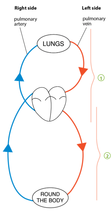
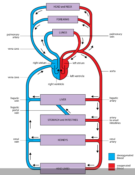
humans and other mammals → Blood passes thru heart twice in one complete circuit blood flows from other parts of the body to the heart → then from the heart it flows to the lungs. then from lungs it flows back to the heart before it is forced out the rest of the body.
- Pulmonary and Systemic circulation*
- from Heart - pulmonary arteries carry blood to the lungs
- oxygen enters blood in the lungs
- oxygenated blood returned to the heart by pulmonary veins
- oxygenated blood leaves the left side of the heart and is distributed by arteries to all parts of the body (Except the lungs)
- Veins -carry blood from all parts of the body back to the right side of the heart
advantages of double circulation
- Blood enters the lungs at a lower pressure*
- Blood flows slowly through the lungs to ensure sufficient time for oxygen absorpton
- heart forced out blood at a higher pressure to the rest of the body
- oxygenated blood is distributed to the rest of the body faster
- oxygenated blood is distributed to the rest of the body faster
The Heart
IMPORTANT: THE HEART
What happens when we sit for too long
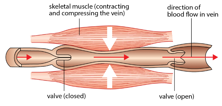
- pressure can build up in the veins of the lower limbs
- causes poor blood circulation and blood to pool in the legs
- Skeletal muscles and movement are important to ensure blood returns to the heart.
Main blood vessels through which heart pumps blood
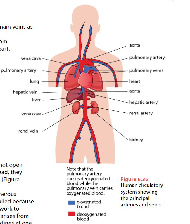
Main arteries leaving body:
- Pulmonary arteries from right ventricle
- Aorta from left ventricle
From aorta, following arteries rise:
- Arteries to the head, neck and arms
- Hepatic artery to liver
- Artery to stomach and intestines
- Renal Arteries → one to each kidney
Main veins in the body:
Blood is returned to the heart by the main veins as follows:
- Pulmonary veins bring blood from lungs to the left atrium of the heart
- Upper Vena Cava returns blood from head, neck and arms to the right atrium
- Lower vena cava returns blood from the rest of the body (excluding lungs) to the right atrium
Lower vena cava receives blood from various veins from lower part of the body:
- Renal Vein bringing blood from Kidneys
- Hepatic vein bringing blood from the liver
Veins from Small Intestine → do not open directly into lower vena cava.
→ they unite to form the hepatic portal vein → which enters the liver and branches into numerous capillaries there.
→ ‘portal vein’ → so called → because it carries blood from one capillary network to another.
→ hepatic portal vein → arises from capillaries in the wall of the small intestine → at one end → and gives rise to capillaries in liver at the other end.
Tissue fluid and lymph ***
- Fluid leaks from blood into surrounding tissues as blood circulates
- nutrients and salts move out into tissues
- fluid is known as lymph/tissue fluid
Formation of tissue fluid:
- Blood pressure at arterial end of capillaries is high
- Blood Plasma → without protein → forced out through capillary walls.
- Contains Phagocytes which are able to squeeze out
- Tissue fluid is collected as lymph → will travel into another system of tubes which returns to the blood.
- these tubes are lymphatic vessels
- Lymphatic vessels empty lymph into the vena cava and eventually the heart.
Coronary Heart Disease
Important
venous end has lower water potential and blood pressure due to loss of water at the arterial end. this causes water to re-enter the capillary at the venous end
Sample
blood clots in brain (or any other part for that matter) can affect brain. Explain.
- the clot causes a blockage which prevents oxygen glucose, amino acids, and other essential substances from reaching the brain cells. this stops (aerobic) respiration and other vital processes from occurring and thus leads to cellular death (energy not released via respiration). thus the part of the brain affected is not able to function.
Sample
small arteries have larger proportion of muscle fibres than large arteries. suggest role for muscle fibres in small arteries
- contraction of muscle fibres helps to push blood forward as pressure exerted from left ventricle reduces with distance from the heart. the muscles can also function to divert blood flow to where it is needed through vasodilation and vasoconstriction
Sample
Describe what causes the decrease in pressure as blood flows from aorta to vena cava
- Decrease in muscle and elastic tissue in the blood vessels from the aorta to the vena cava reduces their ability to exert force required to maintain BP.